Radius Bone Labelled Diagram / Forearm Hand Bones Labeled Diagram Stock Illustration 296617250 : Cheek bone (zygoma) upper jaw (maxilla).. The radius is the home for a few muscles' insertion points. At the humerus, they articulate with the condyle. The radius or radial bone is one of the two large bones of the forearm, the other being the ulna. The skeletal system is a topic of the event anatomy for the 2020 competition, along with the integumentary system and the muscular system. The radius' main functions are to articulate with the ulna and humerus at the elbow to provide supination and pronation.
The side towards your thumb is called lateral and the side towards your little finger is called medial. It provides strength and works with the ulna to allow the wrist and hand to rotate. The radius and ulna are the two bones of the forearm. 12 photos of the labelled diagram of radius bone. The radial head articulates with both the radial notch of the ulna and the humerus at the capitulum and at the trochleocapitellar groove.
Chicken Wing Dissection from www2.nau.edu The tendon of the brachioradialis. Cheek bone (zygoma) upper jaw (maxilla). The skeleton acts as a scaffold by providing support and protection for the soft tissues that make up the rest of the body. Due to the human instinct to break a fall by outstretching the arms, the radius is one of the more frequently fractured bones in the body. It lies laterally and parallel to ulna, the second of the forearm bones. Left human arm is designed based on original size of relevant human bones. Proximal radius includes the cylindrical radial head and neck (figs. Observe and describe the processes of bone remodeling and bone growth.
And radius is often located next to the thumb.
The radius is considered the most commonly fractured bone in the human body, with distal radius fractures being the most common form of radial. Thats the way i remembered which bone is located in anatomy class. Even my bio teacher get confused. The embryonic precursor to both of these is the osteoprogenitor cell, a mesenchymal stem cell. Human body left hand bone images. It is simulated by using a 12 kg/cm servo motor with gears. Following diagram would make it easier to. Learn vocabulary, terms and more with flashcards, games and other study tools. The radius' main functions are to articulate with the ulna and humerus at the elbow to provide supination and pronation. The radius is a long bone in the forearm. There are different features on each bone that also can help distinguish between the. Radius, in anatomy, the outer of the two bones of the forearm when viewed with the palm facing forward. Labelled diagram of radius bone, bone, labelled diagram of radius bone.
Start studying radius bone markings diagram. ( a ) anterior view, ( b ) posterior view. Related posts of labelled diagram of radius bone arms and hands bones names. It extends from the lateral side of the elbow to the thumb side of the wrist and runs parallel to the ulna. At the humerus, they articulate with the condyle.
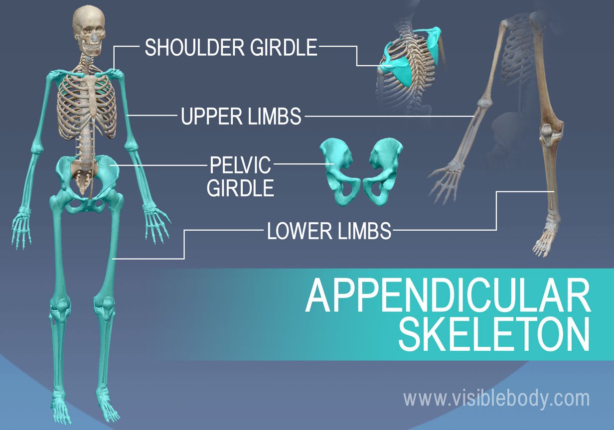
Appendicular Skeleton Learn Skeleton Anatomy from www.visiblebody.com Human body left hand bone images. It's not that clear on this model here, but i'll switch over to another diagram and show you. Each bone is a complex living organ that is made up of many cells, protein fibers, and minerals. The radius bone is the lateral bone of the forearm, and is homologous with the tibia of the lower limb. This articulation occurs in conjunction with the scaphoid. The radius bone is shorter. The embryonic precursor to both of these is the osteoprogenitor cell, a mesenchymal stem cell. For the skeletal system you will need to know:
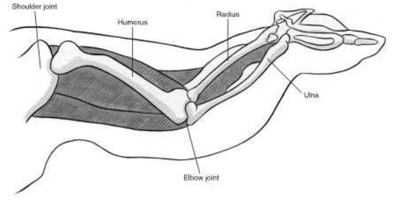
Radius along with ulna connects elbow to forearm. Annotated diagram of the radius and ulna. Skull, clavicle, mandible, scapula, thorax, sternum, humerus, ulna, radius, carpus, phalanges (fingers), metacarpus, spine, pelvis, sacrum, femur, tibia, fibula, tarsus. The ulna articulates with the trochlea and the radius articulates with the capitulum. It is simulated by using a 12 kg/cm servo motor with gears. It is one of the two bones of the forearm, the other being the ulna. Learn radius and ulna anatomy with these fun quizzes and diagrams. The radius' main functions are to articulate with the ulna and humerus at the elbow to provide supination and pronation. The radial head articulates with both the radial notch of the ulna and the humerus at the capitulum and at the trochleocapitellar groove. There are different features on each bone that also can help distinguish between the. All land vertebrates have this bone. The tendon of the brachioradialis. The radius is the home for a few muscles' insertion points.
The radius is the home for a few muscles' insertion points. Left human arm is designed based on original size of relevant human bones. This articulation occurs in conjunction with the scaphoid. Due to that adjacent neural structures may get compressed and produce symptoms of radiculopathy. Lower jaw (mandible) collar bone.
Skeleton Labelled Stock Illustration Illustration Of Graphic 41527284 from thumbs.dreamstime.com It lies laterally and parallel to ulna, the second of the forearm bones. Projection of bone on the lateral surface of the distal radius bone. Start learning now at kenhub. It's not that clear on this model here, but i'll switch over to another diagram and show you. The radius bone is shorter. Radial tuberosity (tuberositas radii) is an oval elevation on the proximal, medioanterior margin of the radius. Annotated diagram of the radius and ulna. The radius bone is the lateral bone of the forearm, and is homologous with the tibia of the lower limb.
The surface opposite to the above mentioned point articulates with the distal part of radius.
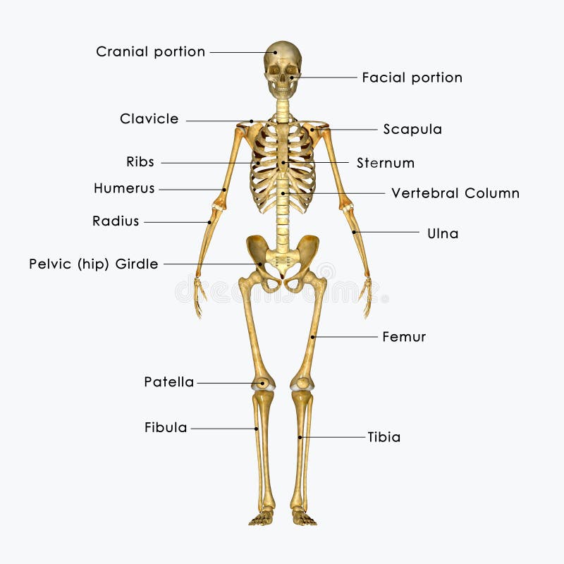
Cheek bone (zygoma) upper jaw (maxilla). Human body left hand bone images. There are different features on each bone that also can help distinguish between the. Following diagram would make it easier to. Skull, clavicle, mandible, scapula, thorax, sternum, humerus, ulna, radius, carpus, phalanges (fingers), metacarpus, spine, pelvis, sacrum, femur, tibia, fibula, tarsus. The shaft is known as the diaphysis and the end of a long bone is called an epiphysis. Left human arm is designed based on original size of relevant human bones. The radius is considered the most commonly fractured bone in the human body, with distal radius fractures being the most common form of radial. All land vertebrates have this bone. Due to that adjacent neural structures may get compressed and produce symptoms of radiculopathy. The bones shown in the chest and hip region in the labeled human skeleton diagram are the ribs humerus is located in the upper arm. It lies laterally and parallel to ulna, the second of the forearm bones. It is one of the two bones of the forearm, the other being the ulna.
Lower jaw (mandible) collar bone labelled radius bone. Projection of bone on the lateral surface of the distal radius bone.


0 Komentar